

EnCor Biotechnology
Mouse Monoclonal Antibody to Microtubule Associated Protein MAP2AB (MAP2), Cat# MCA-4H5
Description
This antibody was raised against purified full length bovine brain MAP2 and the epitope was mapped to amino acids 631-1056 of the human sequence since it bound to EnCor product Prot-r-MAP2-P2, a recombinant human construct continuing these sequences. EnCor markets an antibody which binds all MAP2 isoforms, MCA-2C4, and another mouse monoclonal antibody which binds a different epitope in the projection domain of MAP2A and MAP2B binding epitopes only in MAP2AB MCA-5H11. EnCor also markets popular chicken, rabbit and goat polyclonal antibody recognizing various forms of MAP2 CPCA-MAP2 , RPCA-MAP2ABCD and GPCA-MAP2AB .
- Cell Structure Marker
- Cytoskeletal Marker
- Developmental Marker
- Epitope Mapped Antibodies
- Immunohistochemistry Verified
- Mouse Monoclonal Antibodies
Add a short description for this tabbed section
| Immunogen: | Full length MAP2 purified from bovine spinal cord |
| HGNC Name: | MAP2 |
| UniProt: | P11137 |
| Molecular Weight: | ~280kDa by SDS-PAGE |
| Host: | Mouse |
| Isotype: | IgG1 |
| Species Cross-Reactivity: | Human, Rat, Mouse, Cow |
| RRID: | AB_2572346 |
| Format: | Protein G affinity purified antibody at 1mg/mL in 50% PBS, 50% glycerol plus 5mM NaN3 |
| Applications: | WB, IF/ICC, IHC |
| Recommended Dilutions: | WB: 1:5,000-1:10,000. IF/ICC and IHC: 1:1,000-2,000. |
| Storage: | Store at 4°C for short term, for longer term store at -20°C. Stable for 12 months from date of receipt. |
Microtubules are 25nm diameter protein rods found in most kinds of eukaryotic cells and are associated with a family of proteins called microtubule associated proteins (MAPs). MAPs play a crucial role in the regulation of microtubule dynamics and interactions in vivo. MAP2 was originally named as one of the higher molecular weight MAPs with an SDS-PAGE molecular weight of about 280kDa (1-3). There is a single mammalian MAP2 gene which may generates two high molecular weight proteins of ~280kDa name MAP2A and MAP2B and multiple lower molecular weight forms usually named MAP2C and MAP2D which run on SDS-PAGE gels at 60-70kDa. The lower molecular weight forms are found in neurons early in development, but as the animal matures they are replaced by the higher molecular weight forms (2). The MAP2A and MAP2B forms include a protein sequence which forms fine filamentous protrusions from the sides of brain microtubules, which is therefore referred to as the projection domain. The epitope for this antibody was was mapped to the projection domain sequences so the antibody is specific for MAP2A and MAP2B. This region is one of the prototypes for "intrinsically unstructured regions", a widespread type of protein sequence (4). MAP2 isoforms are expressed only in neurons, specifically in the perikarya and dendrites of these cells. Antibodies to MAP2 isotypes are therefore excellent markers of neuronal dendrites and are useful for identifying neurons in cell culture and sections (e.g. 5-9).

Chromogenic Immunostaining of a 4% PFA fixed paraffin embedded rat hippocampus section with mouse mAb to MAP2, MCA-4H5, dilution 1:1,000,detected with DAB (brown) using the the Vector Labs ImmPRESS method and reagents with citrate buffer retrieval. Hematoxylin (blue) was used as the counterstain. In the hippocampus, MCA-4H5 strongly labels neurons, neuronal projections, and synapses. This antibody performs well in testing with 4% PFA fixed tissues but does not stain NBF fixed human material effectively. We recommend clone MCA-5H11 for human tissue immunohistochemistry. Mouse select image for larger view.

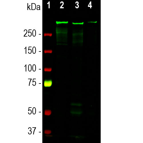
The S lane shows molecular weight standards of the indicated apparent molecular weights in kDa. The next 5 lanes show 2µg amounts of purified recombinant forms of various human MAP2 proteins. D is recombinant full length MAP2D and C is recombinant full length MAP2C. MAP2D is 559 amino acids while MAP2C is 471, missing one of the microtubule binding domains of MAP2D and a more N-terminal 57 amino acid N-terminal sequence. Lanes P1, P2 and P3 show each recombinant segments of the human MAP2 projection domain, found in MAP2A and B but absent from MAP2C and D. The P1 construct is amino acids 233-684, the P2 is 712-1136 and P3 is 1137-1588. All of the constructs run more slowly on SDS-PAGE than expected from their molecular size, probably due to their high content of charged amino acids. These constructs were used to make a series of mouse monoclonal antibodies. The lane marked BSA contains 2µg of bovine serum albumin, another gel standard. Mouse select image for larger view.
1. Dehmelt H, Halpain S. The MAP2/Tau family of microtubule-associated proteins.
Genome Biol. 6:204 (2005).
2. Nunez J. Immature and mature variants of MAP2 and tau proteins and neuronal plasticity. Trends Neurosci. 11:477-9 (1998).
3. Vallee R. A taxol-dependent procedure for the isolation of microtubules and microtubule-associated proteins (MAPs). J. Cell Biol. 92:435-42 (1992).
4. Tompa P. Intrinsically unstructured proteins. Trends Biochem. Sci. 27:527-33 (2002).
5. Goetz AK, et al. Temporally restricted substrate interactions direct fate and specification of neural precursors derived from embryonic stem cells. PNAS 103:11063-8 (2006).
6. Walton, NM, et al. Gliotypic neural stem cells transiently adopt tumorigenic properties during normal differentiation. Stem Cells 27:280-9 (2009).
7. Gasser, A. et al. An ankyrinG-binding motif is necessary and sufficient for targeting Nav1.6 sodium channels to axon initial segments and nodes of Ranvier. J. Neurosci. 32:7232-43 (2012).
8. Rush AM, et al. Differential modulation of sodium channel Nav1.6 by two members of the fibroblast growth factor homologous factor 2 subfamily. Eur. J. Neurosci. 23:2551-62 (2006).
9. Eckenstein FP, McGovern T, Kern D, Deignan J. Neuronal vulnerability in transgenic mice expressing an inducible dominant-negative FGF receptor. Exp. Neurol. 198:338-49 (2006).
Add a short description for this tabbed section


,%20Cat%23%20MCA-4H5)