

EnCor Biotechnology
Mouse Monoclonal Antibody to Myelin Basic Protein (MBP), Cat# MCA-7D2
Description
The MCA-7D2 antibody was made against a preparation of MBP purified biochemically from bovine brain. It can be used to identify oligodendrocytes and Schwann cells in neural cell culture, to visualize myelin sheaths and myelinating cells in sectioned material and to probe western blots for MBP gene products. The antibody is also rather insensitive to aldehyde fixation and so can be used in immunohistochemistry of paraffin sections and in CLARITY type applications. The MCA-7D2 antibody binds only the 21.5kDa and 18.5kDa rat MBP isotypes, but all four isotypes of human and bovine MBP, mapping the epitope to amino acids 145-184 of the human 21.5kDa sequence. The Kon rate is 2.189 x 10⁻⁵, the Koff rate is 4.86 x 10⁻⁴ and the Kd is 2.22 x 10⁻⁹. The antibody works well for western blotting and for IF, ICC and IHC (for IHC see data under "Additional Data" tab). Our alternate mouse monoclonal MCA-7G7 binds to all four rat MBP gene products, mapping the epitope to the "core" of the MBP protein. A sequence alignment of the four CNS MBP isotypes in human and rat can be downloaded from here. We also generated chicken and goat polylconal antibodies to MBP, CPCA-MBP and GPCA-MBP.
- Cell Structure Marker
- Cell Type Marker
- Epitope Mapped Antibodies
- Immunohistochemistry Verified
- Mouse Monoclonal Antibodies
Add a short description for this tabbed section
| Immunogen: | Purified myelin basic protein isolated from bovine brain, epitope maps to the peptide AEGQRPGFGYGGRASDYKSAHKGFKGVDAQGTLSKIFKLG, amino acids 145-184 of the human 21.5kDa sequence. |
| HGNC Name: | MBP |
| UniProt: | P02687 |
| Molecular Weight: | 18.5 and 21.5kDa human isotypes |
| Host: | Mouse |
| Isotype: | IgG1 |
| Species Cross-Reactivity: | Human, rat, mouse, cow, pig, horse |
| RRID: | AB_2140350 |
| Format: | Protein G affinity purified antibody at 1mg/mL in 50% PBS, 50% glycerol plus 5mM NaN3 |
| Applications: | WB, IF/ICC, IHC |
| Recommended Dilutions: | WB: 1:5,000-1:10,000. IF/ICC: 1:2,000-5,000. IHC: 1:10,000. |
| Storage: | Store at 4°C for short term, for longer term store at -20°C. Stable for 12 months from date of receipt. |
Myelin Basic Protein (MBP) is one of the major proteins of the myelin sheath surrounding axons in the nervous system. The protein sequence was determined from purified protein over 30 years ago (1). The protein is made by oligodendrocytes in the central and nervous system, so antibodies to MBP are good markers of this cell type. In the peripheral nervous system MBP is expressed by myelinating Schwann cells so this antibody can be used to identify these cells in culture or sections. In the central nervous system four different forms of the protein made by alternate transcription from a single gene, the protein products having molecular weights of 21.5, 20.5, 18.5, and 17.2kDa in humans. The single gene of rodents also produces 4 different proteins, but of slightly different sizes, 21.5, 18.5, 17 and 14kDa. Some interest has focused on MBP as a potentially significant auto-antigen involved in mouse models of multiple sclerosis (MS, 3) and in human patients (4). Detection of MBP released into blood and CSF has some potential as a surrogate biomarker of demyelination and axonal loss in MS and other damage and disease states (e.g. 5).

Chromogenic immunostaining of a formalin fixed paraffin embedded human brain cortex section with mouse mAb to MBP, MCA-7D2, dilution 1:10,000, detected with DAB (brown) using the the Vector Labs ImmPRESS method and reagents with citrate buffer retrieval. Hematoxylin (blue) was used as the counterstain. The MBP antibody labels axonal myelin sheaths. This antibody performs well in testing with both 4% PFA and standard NBF fixed rat and human tissues. Mouse select image for larger view.

Immunofluorescent analysis rat brain cerebellum section stained with mouse mAb to Myelin Basic Protein (MBP), MCA-7D2, dilution 1:5,000 in green, and costained with rabbit pAb to NF-M, RPCA-NF-M, dilution 1:2,000 in red. The MBP antibody stains myelin sheathes around axons, while the NF-M antibody labels dendrites and axons of neuronal cells. Mouse select image for larger view.

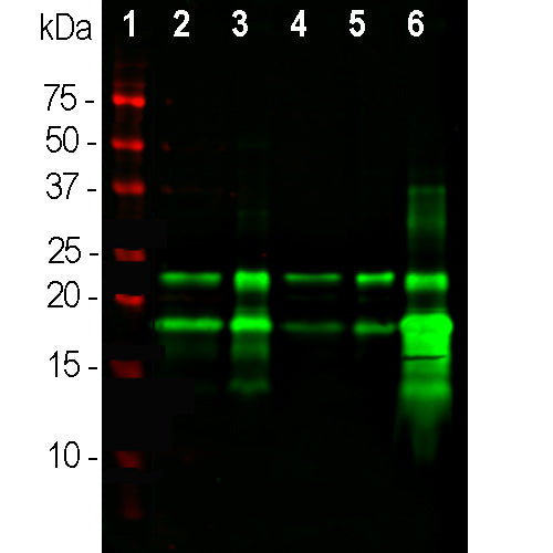
Blots of 20µg crude rat brain homogenate were probed with 2 MBP antibodies; MCA-7G7 (left lane) at 1:5,000 and MCA-7D2 (right lane) at 1:5,000 dilution. MCA-7D2 binds the largest 21.5kDa and 18.5kDa isotypes preferentially, while monoclonal MCA-7G7 binds all four transcripts running at 21.5, 18.5, 17 and 14kDa. Position of nmolecular weight standards are at left side at indicated size in kDa. Mouse select image for larger view.
To download a sequence alignment of the 4 human and rat MBP isotypes link to here.
1. Eylar EH, et al. Basic A1 protein of the myelin membrane. The complete amino acid sequence. J. Biol. Chem. 246:5770-84 (1971).
2. Marty MC, et al. The myelin basic protein gene is expressed in differentiated blood cell lineages and in hemopoietic progenitors. PNAS 99:8856-61 (2002).
3. Libbey JE, Fujinami RS. Experimental Autoimmune Encephalomyelitis as a Testing Paradigm for Adjuvants and Vaccines. Vaccine 29:3356–62 (2011).
4. Wucherpfennig KW, Strominger JL. Molecular mimicry in T cell-mediated autoimmunity: Viral peptides activate human T cell clones specific for myelin basic protein. Cell 80:695-705 (1995).
5. Berger RP, et al. Serum neuron-specific enolase, S100B, and myelin basic protein concentrations after inflicted and noninflicted traumatic brain injury in children. J. Neurosurg. 103:61-8 (2005).
Add a short description for this tabbed section


,%20Cat%23%20MCA-7D2)