

EnCor Biotechnology
Mouse Monoclonal Antibody to Neurofilament NF-L (Nfl, NEFL), Cat# MCA-7D1
Description
MCA-7D1 was made against a preparation of full length native NF-L protein purified from porcine spinal cord. It binds NF-L from a variety of species including human, rat and mouse. The epitope is not fully characterized since it could not be localized using 20 amino acid nested peptides but is known to be not within the α-helical region, amino acids 88-400 of the human protein. The antibody works well for WB, IF, ICC and IHC. We also market several other NF-L antibodies including rabbit and chicken polyclonal antibodies RPCA-NF-L and CPCA-NF-L.
- Cell Structure Marker
- Cell Type Marker
- Cytoskeletal Marker
- Mouse Monoclonal Antibodies
- Not Recommended for IHC
Add a short description for this tabbed section
| Immunogen: | Full length native protein purified from pig spinal cord |
| HGNC Name: | NEFL |
| UniProt: | P07196 |
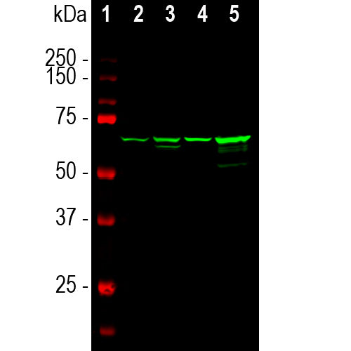
| Molecular Weight: | 68-70kDa |
| Host: | Mouse |
| Isotype: | IgG2b heavy, κ light |
| Species Cross-Reactivity: | Human, rat, mouse, cow, pig, horse |
| RRID: | AB_2572363 |
| Format: | Protein G affinity purified antibody at 1mg/mL in 50% PBS, 50% glycerol plus 5mM NaN3 |
| Applications: | WB, IF/ICC, IHC |
| Recommended Dilutions: | WB: 1:2,000-1:5000. IF/ICC, and IHC: 1:1000-1:5000. |
| Storage: | Store at 4°C for short term, for longer term store at -20°C. Stable for 12 months from date of receipt. |
Neurofilaments are the 10nm or intermediate filament proteins found specifically in neurons, and are composed predominantly of three major proteins called NF-L, NF-M and NF-H, though other filament proteins may be included also. The major function of neurofilaments is likely to control the diameter of large axons (1). NF-L is the neurofilament light or low molecular weight polypeptide and runs on SDS-PAGE gels at 68-70kDa with some variability across species. Antibodies to NF-L like MCA-7D1 are useful for identifying neuronal cells and their processes in cell culture and sectioned material. NF-L antibody can also be useful for the visualization of neurofilament rich accumulations seen in many neurological diseases, such as Lou Gehrig’s disease (ALS), giant axon neuropathy, Charcot-Marie Tooth disease and others (2-4). Much interest has recently been focused on the detection of NF-L released from neurons into blood and CSF as a surrogate marker of primarily axonal loss in a variety of types of CNS injury and degeneration (5).

Chromogenic immunostaining of a formalin fixed paraffin embedded rat cerebellum section with mouse mAb to NF-L, MCA-7D1, dilution 1:1,000, detected with DAB (brown) using the the Vector Labs ImmPRESS method and reagents with citrate buffer retrieval. Hematoxylin (blue) was used as the counterstain. The MCA-7D1 antibody labels neuronal perikarya and their processes. This antibody performs well in testing with both 4% PFA and standard NBF fixed tissues. Mouse select image for larger view.
1. Hoffman et al. Neurofilament gene expression:a major determinant of axonal caliber. PNAS 84:3472-6 (1987).
2. Perrot R, et al. Review of the Multiple Aspects of Neurofilament Functions, and their Possible Contribution to Neurodegeneration. Mol. Neurobiol. 38:27-65 (2008).
3. Lépinoux-Chambaud C. Eyer J. Review on intermediate filaments of the nervous system and their pathological alterations. Histochem. Cell Biol. 140:13-22 (2013).
4. Liu Q. et al. Neurofilamentopathy in Neurodegenerative Diseases. Open Neurol. J. 5:58–62 (2011).
5. Bacioglu M, et al. Neurofilament light chain in blood and CSF as marker of disease progression in mouse models and in neurodegenerative diseases. Neuron 91:56-66 (2016).
Add a short description for this tabbed section


,%20Cat%23%20MCA-7D1)