August 2023 News

      We release yet more novel antibodies. We made a novel rabbit polyclonal and a mouse mpnoclonal to the enzyme catalase, RPCA-Catalase and MCA-6H14, both good markers odf peroxisomes. We also raised rabbit polyclonal antibodies against human Sigma-1 receptor RPCA-Sigma-1r and human γ-synuclein, RPCA-SNCG. We document that all work well on western blots for IF, ICC and IHC.

April 2023 News

      Our recent work on the Uman NF-L antibodies published in pre-print form in BioRχiv was cited for the first time, in JAMA (the Journal of the American Medical Association), only the world’s highest impact medical journal! For a download see here. This work is now published in peer reviewed and freely downloadable form from the journal Brain Communications (https://doi.org/10.1093/braincomms/fcad067). The paper is now also freely downloadable directly from Pubmed https://pubmed.ncbi.nlm.nih.gov/37091583/. We also made a short video Youtube presentation on this work, see here. If you want to check out research utilizing NF-L as a biomarker search for “(NF-L or Nfl or NEFL or “Neurofilament Light”) and biomarker”, over 2,400 publications to date.

      We have now tested all of our current mouse monoclonal antibodies on formalin fixed paraffin embedded sections for standard immunohistochemistry (IHC) of rodent and human tissues. As expected, not every antibody worked in this format, though a significant number did. We show images of each antibody which worked and details of how we did the staining. In the near future e will also add a tag to indicated which of our antibodies are not recommended for use in this format. Why did certain antibodies not work? The reason is because antibodies bind to their targets by protein-protein interactions involving individual amino acids. Formalin fixation for IHC is typically quite extensive and works by modifying amino acid residues some of which may be part of a particular antibody binding site. As a result a subset of monoclonal antibodies are expected to have reduced or no ability to bind material processed for IHC. We routinely use antigen retrieval methods as this can in many cases reverse the fixation induced antigenic changes, and details of how we obtained each image are outlined on the web pages. Here are some examples, in each case mouse select the image for a larger view.

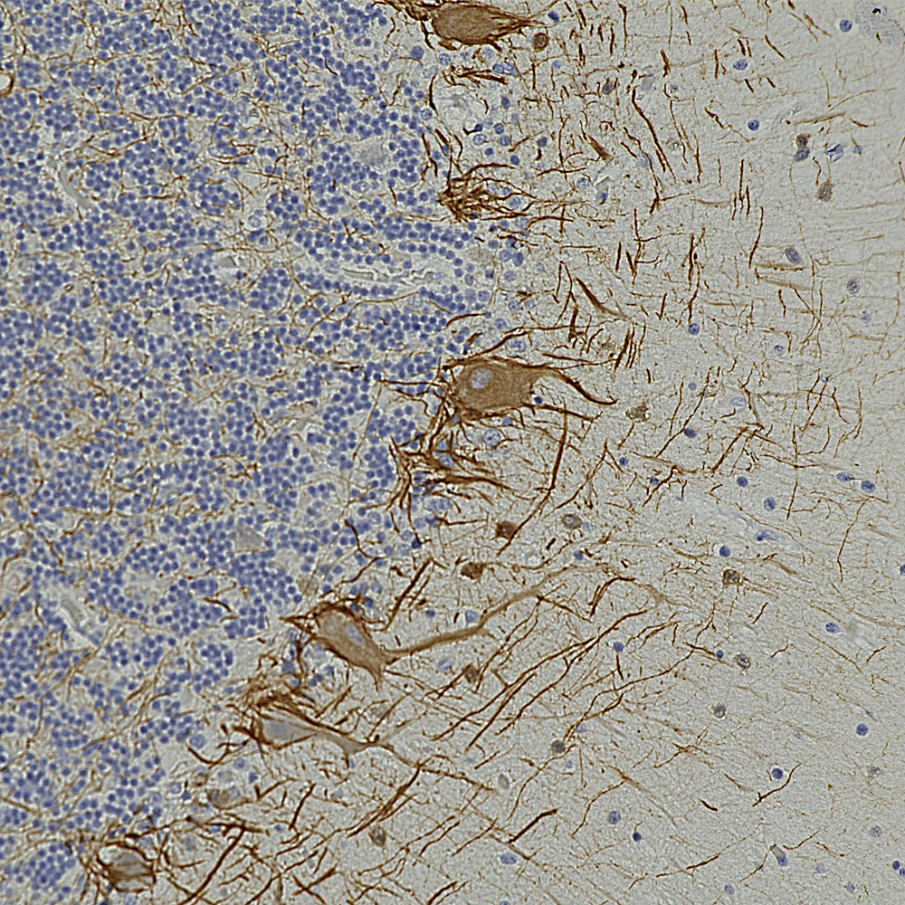
      The image below shows a region of human cerebellum stained with MCA-6H112, an epitope mapped antibody raised against the C-terminal peptide of human neurofilament light chain (NF-L) which reveals basket cell processes, parallel fibers and other kinds of axon in both the molecular and granular layers. A version of this image has been posted on Wikipedia for general use, see here.
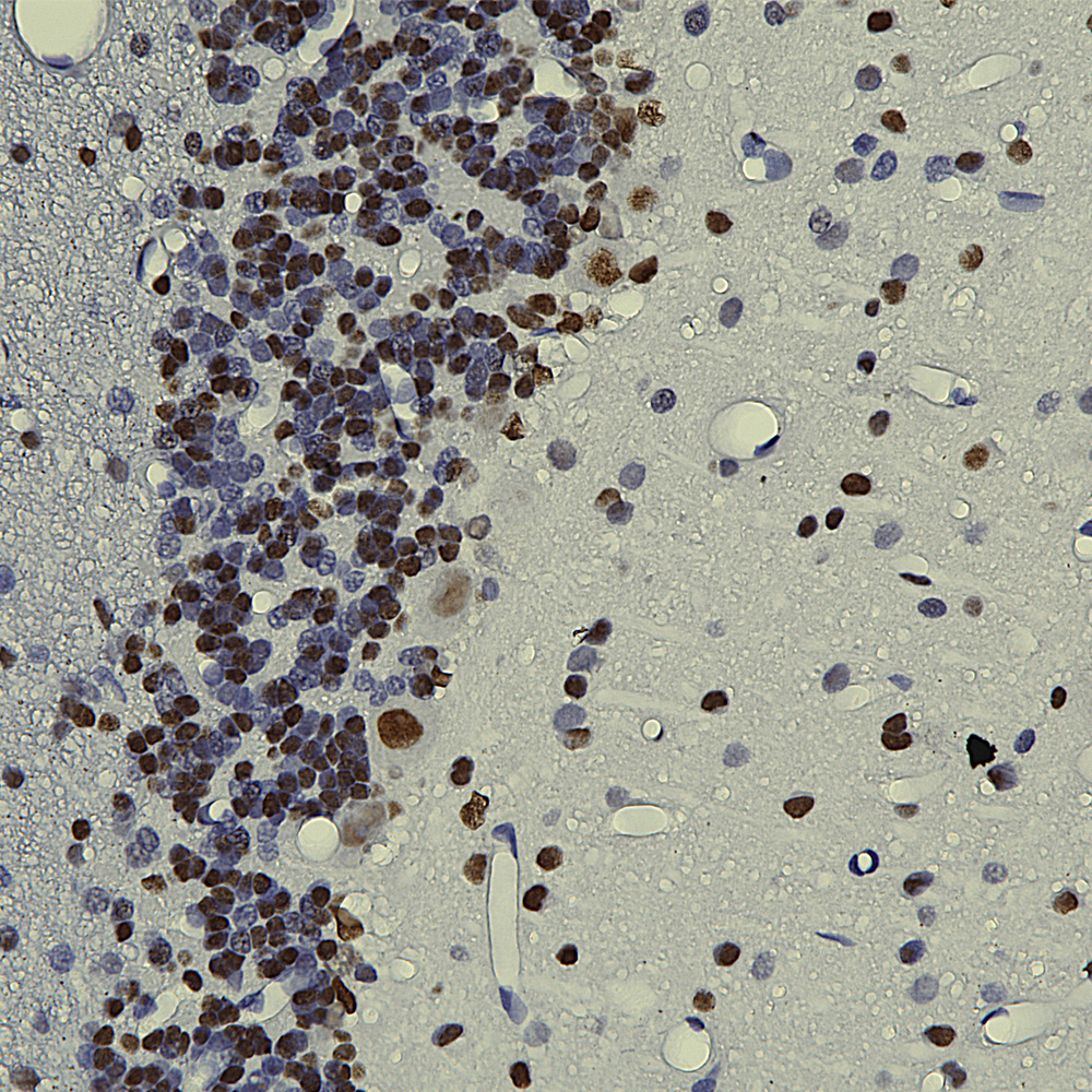
      Below is an IHC image of rat cerebellum showing neuronal nuclear staining of with MCA-3H8, our mouse monoclonal antibody to TDP43. TDP43 accumulates in the nuclei of Purkinje cells and other cerebellar neurons.
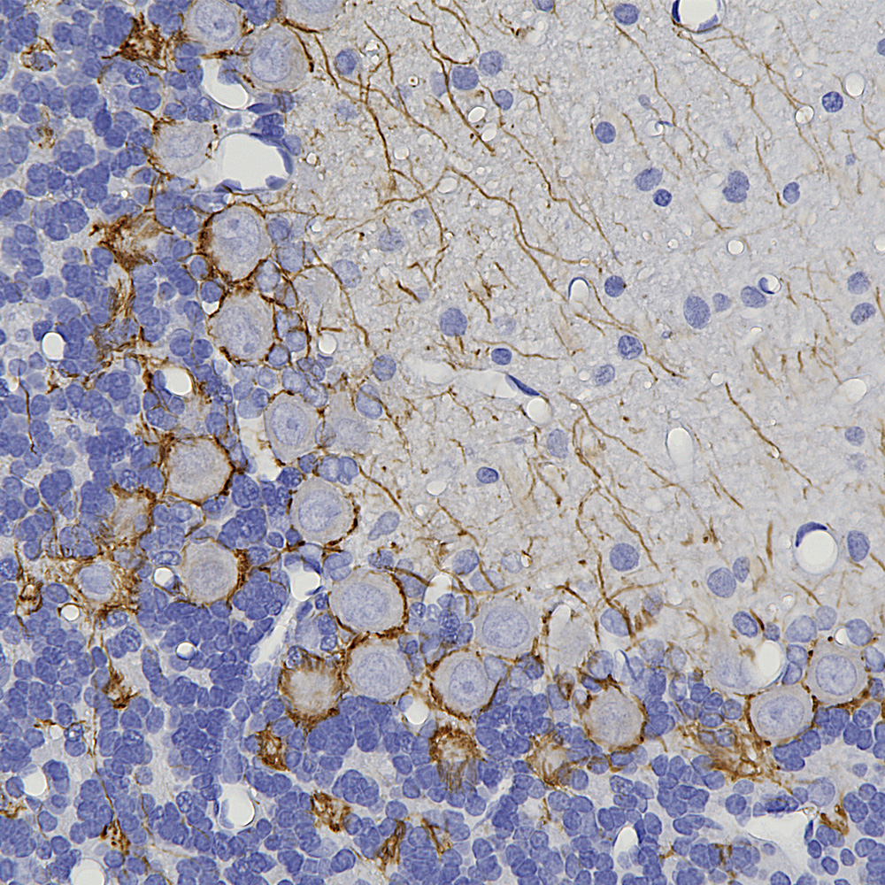
      Below is an example of rat cerebellum stained with MCA-3H11, our eoitope mapped mouse monoclonal antibody to neurofilament medium (NF-M), showing basket cell process and parallel fibers.
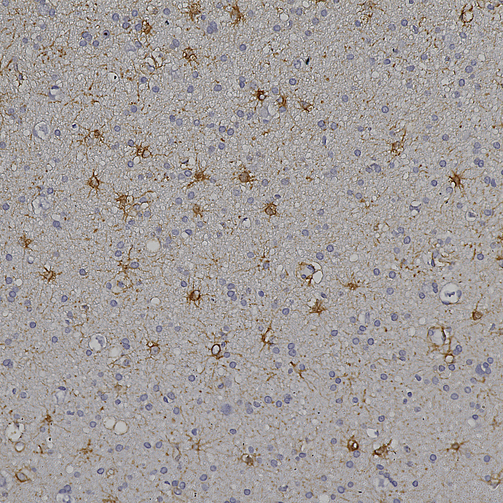
      Below is an example of formalin fixed paraffin embedded human cerebral cortex stained with our mouse monoclonal MCA-4A12 to ALDH1-L1, a marker of astrocytes. This antibody produces a different image of astrocytes than that revealed by GFAP antibodies, since ALDH1-L1 is found throughout the cytoplasm of astrocytes while GFAP reveals the cytoskeletal structural core of these cells.
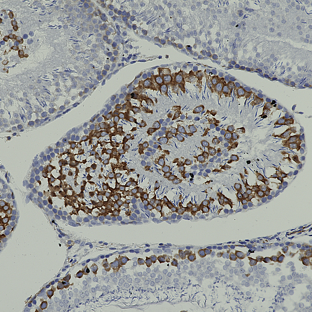
      And below is an example of IHC of rat testis stained with our antibody to Nestin, MCA-4D11.
      

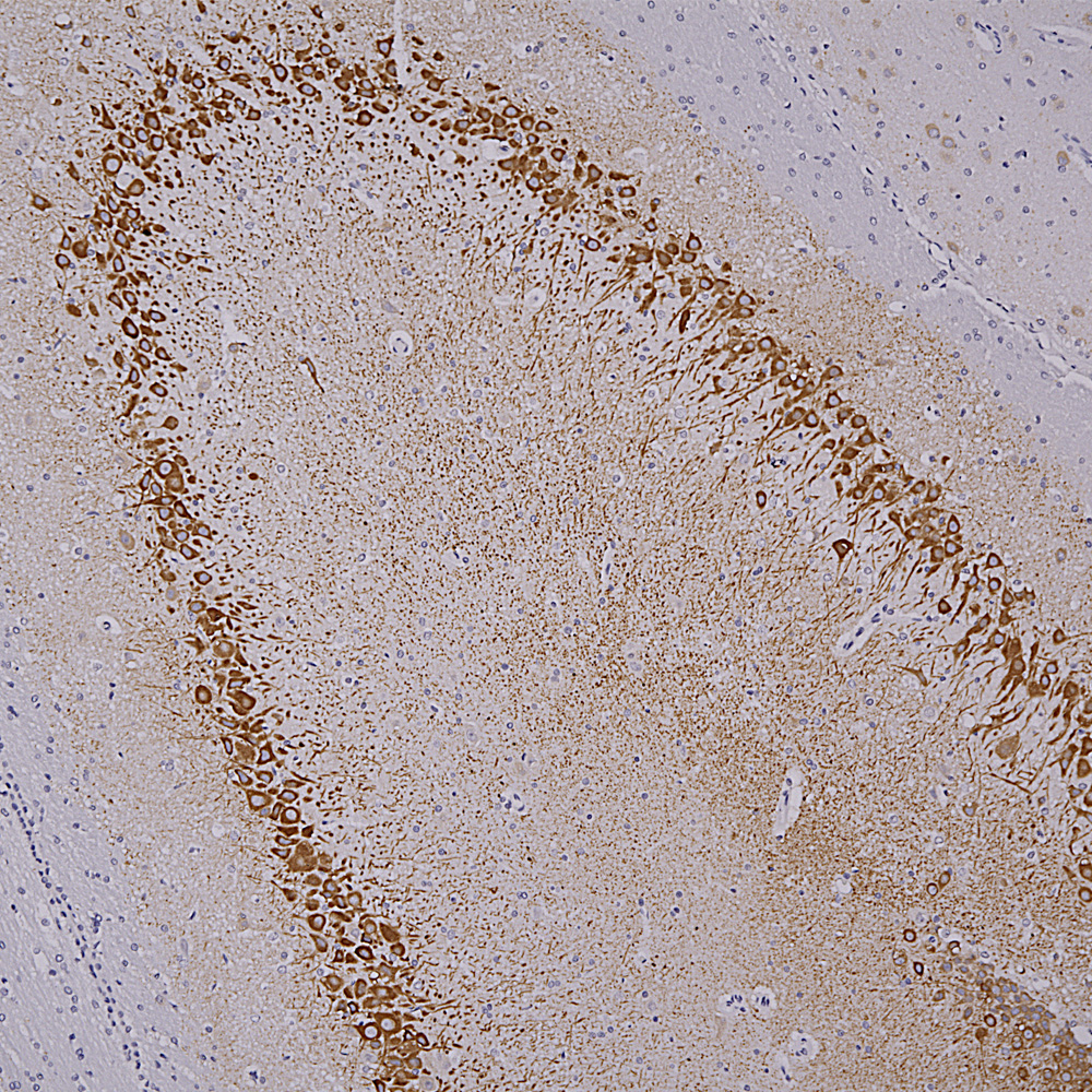
      Below is rat hippocampus stained with MCA-4H5 one of our MAP2 antibodies showing staining for hippocampal pyramidal cells.
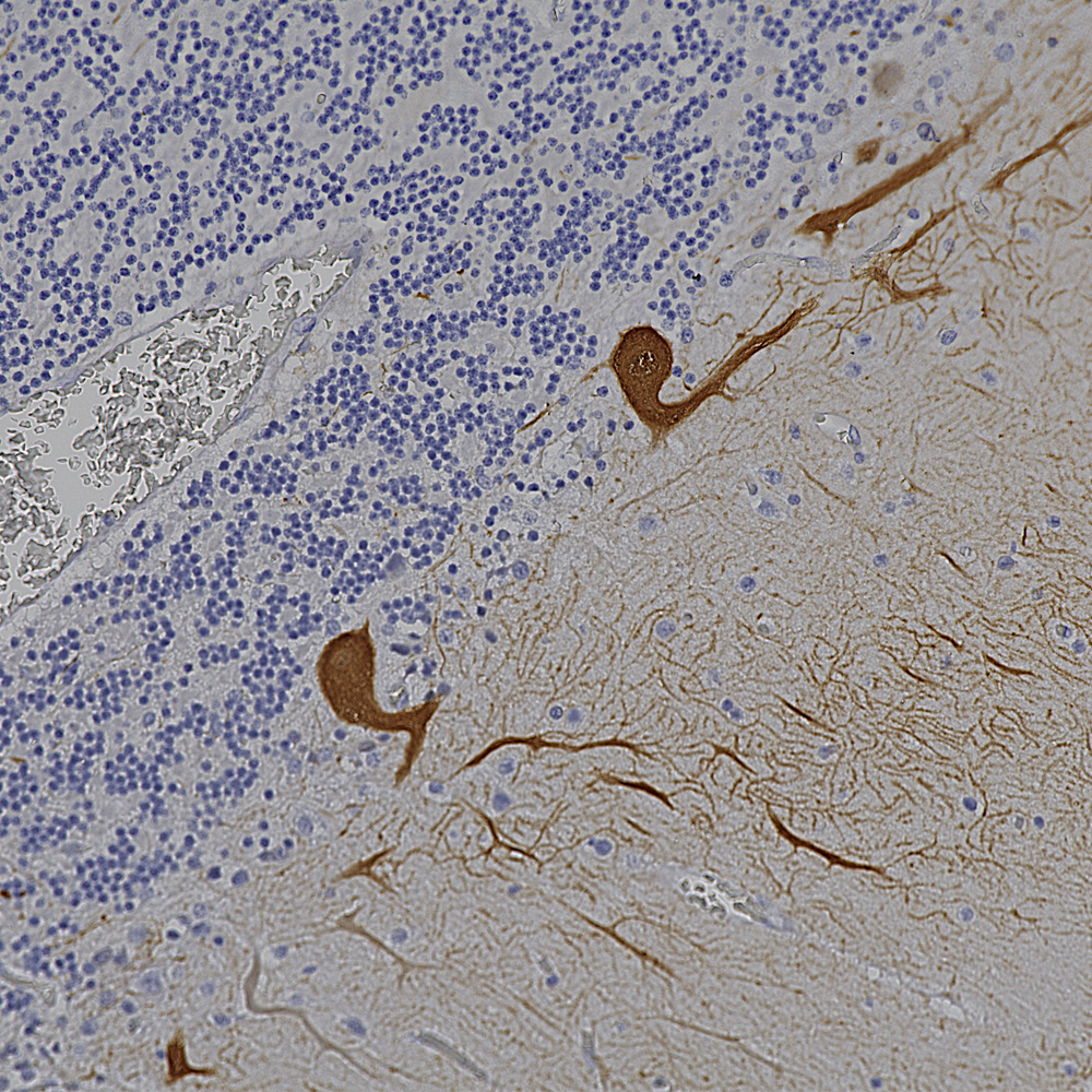
      Below is an IHC specimen of human cerebellum stained with MCA-4H7 mouse monoclonal antibody to calbindin which reveals Purkinke cell dendrites in the molecular layer.
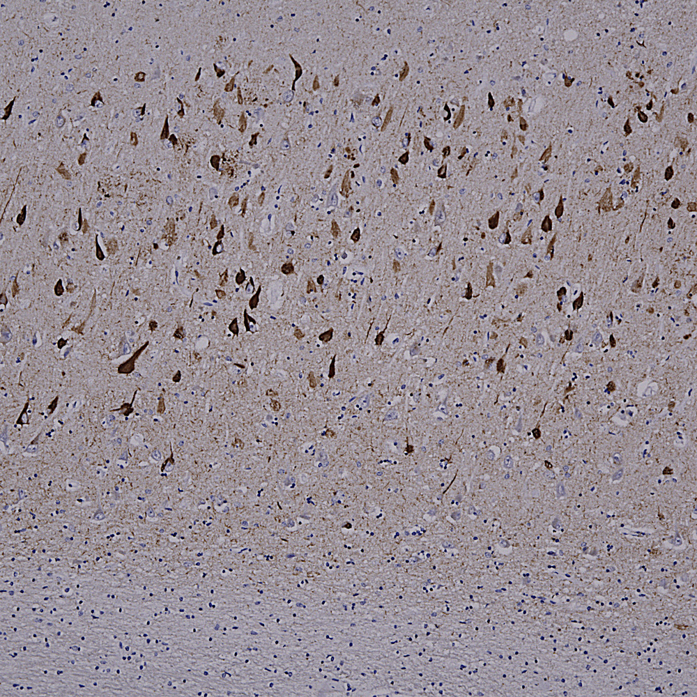
      Below is an example of IHC staining with MCA-5B10, our epitope mapped monoclonal antibody to MAP-τ showing flame shaped tangles in the hippocampus of an Alzheimer’s disease patient.
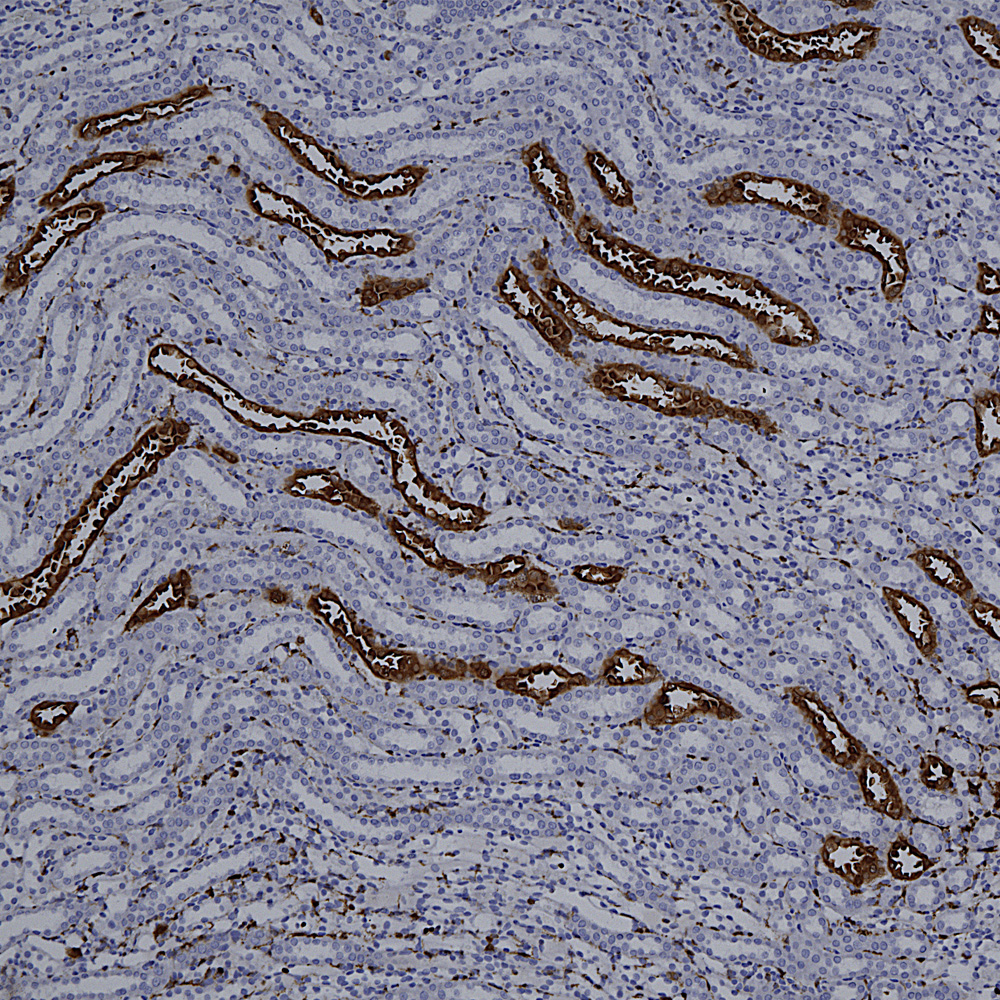
      Below is a section of rat kidney stained with MCA-5C21, our monoclonal antibody to Galectin 3.
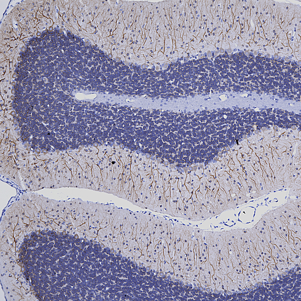
      Below is IHC of rat cerebellum stained with our monoclonal antibody to MAP2 MCA-5H11, showing prominent Purkinje cell bodies and dendrites in the molecular layer.
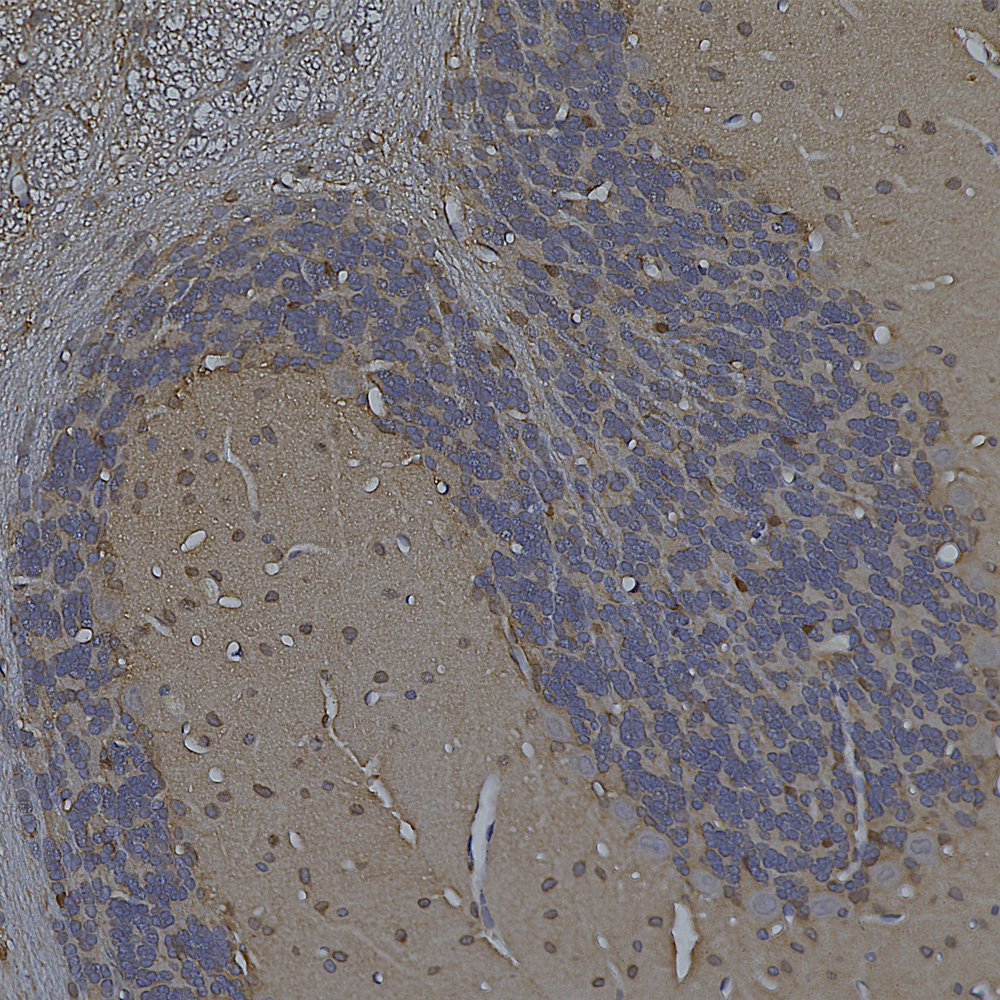
      Finally here is IHC of rat cerebellum stained with our MCA-253 monoclonal to non-neuronal enolase, also known as Eno1 or α-enolase, which stains astrocytes and other non-neuronal cells.
March 2023 News

      The data in our recent publication in BioRχiv preprint, entitled “Uman Type NF-L Antibodies Are Effective Reagents for the Imaging of Neurodegeneration” was edited and modified to include further data and was submitted for peer review at the journal “Brain Communications“. It has now been accepted for publication with the same title except that “NF-L” is replaced with “Neurofilament Light” in the title, we added some more data and also another coauthor. An unedited and unformatted version of the paper appeared on-line, see 10.1093/braincomms/fcad067. The same link will access the edited and formatted version of the paper which will be available soon.
      We also add new data for many of our mouse monoclonal antibodies which we have now screen for immunohistochemistry (IHC) on paraffin embedded and formalin fixed human and rodent tissues. For example our widely used mouse monoclonal antibody to c-FOS, MCA-2H2, works well on rat hippocampus highlighting a few active neurons. These cells were expressing large amounts of c-FOS and therefore activating transcription of other genes at the time the tissue was processed and with current techniques can be characterized in terms of mRNA expression and connections. This data is shown under the “Additional Info” tag on the MCA-2H2 web page. Many of our antibodies perform very well and we have posted images under the “Additional Info” tag on the relevant web pages. As expected a few antibodies did not work well and we will add “not recommended for IHC” on the web page, alonfg with details of a n alternate reagent idf we have one. So far we have data on MCA-1B7, MCA-1C51, MCA-2H2, MCA-1C7, MCA-1B11, MCA-1C7, MCA-1C51, MCA-1D2, MCA-1D4, MCA-1C7, MCA-1E3, MCA-1F1, MCA-1H10, MCA-2A7 and MCA-2A8. We are currently screening our entire panel of mouse monoclonal antibodies for utility in IHC, and will shortly post the rest of our results of this.

August 2022 News

      The Uman Diagnostics NF-LIGHT™ ELISA is a widely used ELISA type assay based on two mouse monoclonal antibodies to the neurofilament NF-L protein. One Uman antibody called variously UD1 or 2.1 is the detection reagent and the other, UD2, also known as 47.3, is the capture reagent (see Norgren et al. 2002). The same pair of antibodies are used in the more sensitive “Single Molecule Array” (Simoa™) assay marketed by Quanterix. These NF-L based assays have become very informative for the quantitation of axonal loss associated with a variety of CNS damage and disease states, see for example Barro et al. 2020 and Gaetani et al. 2019. We now have fully characterized the epitopes for both antibodies and have deposited the basic data on a preprint server see BioRχiv 2022.08.27.504533v1. The data in this report is currently undergoing peer review. These findings greatly increase understanding of what the Uman NF-LIGHT™ ELISA and the Quanterix Simoa™ assay are actually measuring and sets the stage for further NF-L assays with possibly improved properties.
      To our great surprise, neither Uman antibody stained sections of healthy CNS tissue with a typical NF-L pattern. However axons which were damaged as a result of experimental spinal cord injury in rats were strongly reactive with both Uman reagents. We hypothesized that the Uman epitopes were masked in assembled neurofilaments and made available to antibody binding by degeneration induced proteolysis. In agreement with this hypothesis we could make previously Uman negative control tissues strongly Uman positive by treatment with proteases. In addition fresh CNS tissues did not stain with Uman reagents while tissues left to sit at room temperature for 4 hours were strongly reactive. We also discovered that our antibodies to the C-terminal of NF-L, such as our rabbit polyclonal RPCA-NF-L-ct and mouse monoclonal MCA-DA2 stained neurofilaments in healthy processes but generally did not stain the degraded Uman positive NF-L positive material. On closer examination we determined that during injury induced degeneration processes positive for the C-terminal of NF-L became swollen and beaded. They apparently start to express the Uman epitopes and lose the NF-L C-terminal epitopes at about the same time. The final product is mostly diffuse and globular Uman positive degenerated axonal material. The Uman reagents and our NF-L C-terminal antibodies can therefore be used to positively identify both healthy, degenerating and degenerated processes.
      Based on these findings we made a novel panel of antibodies, both monoclonal and polyclonal, to the peptide containing both Uman epitopes. We have named these “DegenoTag™” reagents, as they specifically identify degenerated processes. These have the same properties of the Uman reagents as described above. We therefore have a panel of well characterized antibodies which can be used in the development of ELISA reagents and in immunocytochemical studies of normal, damaged and diseased CNS tissues. The first of these are MCA-6H63, MCA-1B11 and MCA-1D44. MCA-6H63 has an epitope a few amino acids N-terminal to that of UD2 while the epitope for MCA-1D44 is very similar to that of UD2. MCA-1B11 has an epitope similar to that of UD1. MCA-1D44 and MCA-6H63 share the interesting Uman property of only binding to degenerating and degenerated cells and processes, while MCA-1B11 also stains this degenerated material strongly but shows some reactivity with undamaged neurons and processes. We have also developed a rabbit polyclonal antibody RPCA-NF-L-Degen to the same region of NF-L which shares the property of only recognizing forms of NF-L expressed in degenerating processes.
      We publish another research report in collaboration with scientists at the University of Florida. This is “DAT and TH expression marks Human Parkinson’s Disease in Peripheral Immune Cells” by Gopinath et al. from the lab of Habibeh Khoshbouei in the journal “npj Parkinson’s Disease”. This article make use of our tyrosine hydroxylase (TH) antibodies to study blood cells in Parkinson’s disease patients. We also release an excellent and novel goat polyclonal antibody to tyrosine hydroxylase GPCA-TH.

August 2020 News

     We add another mouse monoclonal antibody to the astrocytic and biomarker protein GFAP, MCA-3E10. This antibody was made against recombinant human GFAP and the epitope and kinetics of this antibody are known. It has utility as a component of assays designed to detect GFAP in blood, plasma and sera, which has become of potential interest as a surrogate marker of CNS damage and disease. We also release a chicken polyclonal antibody to neuron specific enolase, a.k.a. enolase 2, CPCA-NSE and a rabbit antibody to fatty acid binding protein 7, RPCA-FABP7. Finally we market two new antibodies to annexin A6, a useful marker of cellular membrane damage and “blebs”, herniations of the cell membrane associated with apoptosis. These are mouse monoclonal MCA-4G3 and rabbit polyclonal RPCA-ANXA6.

July 2020 News

      Like many other laboratories with expertise with antibody production we have focused some urgent attention on the SARS-Cov2 virus and the ACE2 protein which it uses to enter and infect human cells. The region of the virus spike or S-protein and the corresponding virus binding region on the ACE2 protein were recently characterized so we made recombinant forms of both regions in E. coli (Prot-r-SARS-CoV2-bd and Prot-r-ACE2-bd). We now have a panel of excellent mouse monoclonal antibodies to the ACE2 binding site of the SARS-CoV2 S-protein, and are marketing the first two of these, MCA-5G8 and MCA-2G1. We also made an affinity purified rabbit polyclonal antibody raised against the same immunogen, RPCA-SARS-CoV2-bd. These antibodies can be used to identify the SARS-Cov2 virus on western blots and by immunofluorescence and may also block the binding of the virus to ACE2. Of general interest to the world, the SARS-CoV2 S-protein ACE2 binding region turns out to be an excellent immunogen, so that it is likely an ideal target for vaccine development.
      We release new antibodies to β-synuclein, a mouse monoclonal and a rabbit polyclonal, MCA-6A10 and RPCA-SNCB. β-synuclein, like the closely related α-synuclein, is a major brain protein concentrated in synaptic regions. We also release new antibodies to Annexin A5 MCA-6A12 and RPCA-ANXA5. Annexin A5 is a Calcium and membrane associated protein which is upregulated when cells become apoptotic. We also release two new mouse monoclonals directed against PEA-15 MCA-4D2 and MCA-4D117. This protein was discovered as a “phosphoprotein enriched in astrocytes of 15kDa” (PEA-15) and independently as a “protein enriched in diabetes” (PED) and so is sometimes referred to as PED/PEA-15. PEA-15 is one of the large family of proteins containing a death effector domain (DED) and appears to have important functions in the regulation of cell growth, apoptosis and glucose metabolism. Finally we release a goat polyclonal antibody to myelin basic protein, GPCA-MBP, which can be used to identify and study myelin sheathes, oligodendrocytes and certain types of Schwann cell.

December 2019 News

      We have added more quantitative data about our various monoclonal antibodies, including several of the most widely used including our mouse monoclonals to fibrillarin (MCA-38F3 and MCA-4A4), also HSP60, (MCA-1C7), TDP43 (MCA-3H8) and several others. We also have new data on our widely used neurofilament NF-L antibodies (MCA-DA2 and MCA-7D1) and also phosphorylated neurofilament H (MCA-NAP4, MCA-AH1 and MCA-9B12). All this new data will be added to the relevant web pages and data sheets over the next few days.

October 2019 News

We are going to the 2019 Society for Neuroscience meeting in Chicago, so come by Booth 468 to get some pens, flashlights, posters and other free stuffs! We also continue to improve the characterization of our antibodies, so we now know both the epitopes and the kinetic properties of an increasing number, for example see the latest data on our popular mouse monoclonals to FOX3/NeuN, MCA-1B7, cFos, MCA-2H2 and myelin basic protein, MCA-7D2. We also release a new mouse monoclonal to neurofilament NF-L MCA-6H112 directed against the C-terminal peptide of this major structural protein. This antibody works well on blots, cell culture, sectioned material and in CLARITY and related tissue clearing methods, revealing neurons and their axons, and can also be used to monitor neurofilament proteolysis.

April 2019 News

      We add a novel chicken polyclonal antibody which recognizes the neuron specific enolase (NSE), a.k.a. enolase 2 or γ enolase, CPCA-NSE. This recognizes NSE cleanly on western blots and binds neurons and their processes in immunofluorescense experiments. This is a complement to our rabbit polyclonal antibody to NSE, RPCA-NSE and our epitope mapped and highly specific mouse monoclonal antibody to the related protein enolase 1, also known as non-neuronal enolase or α enolase MCA-253.